


Arthrosis is a chronic pathology aimed at damage to the joint structures of the musculoskeletal system. The main cause leading to a chronic disease is a metabolic imbalance that leads to a progressive process of a degenerative-dystrophic nature. The targets of the damaging reaction are articular cartilage, connective tissue, bursae, tendons, bones and muscular corset. In the chronic form of the pathology, periarticular muscles participate in the inflammatory process, losing anatomical elasticity due to joint deformation and swelling. In order to eliminate the complications associated with blocking the biomotility of the skeleton, and not to become disabled, you should arm yourself with information about arthrosis - what it is, what are the causes, symptoms and treatment.
Causes and risk factors for the development of pathology
The inflammatory-destructive process in the joints often begins without a reason. Idiopathic (primary) arthrosis has this beginning. The mechanism of development of secondary arthrosis begins after certain conditions and factors, namely:
- Joint injury (fracture, meniscus damage, ligament tear, sprain, compression + contusion, bone fracture).
- Dysplasia (abnormal intrauterine development of joint components).
- Violation of material metabolism.
- Autoimmune-type pathologies (rheumatoid arthritis, psoriasis, autoimmune toxic goiter, systemic lupus erythematosus).
- Nonspecific destructive arthritis (with a purulent component).
- Infections of different etiology (tuberculosis, meningitis, encephalitis, gonorrhea, syphilis, hepatitis).
- Pathologies of the endocrine glands (diabetes mellitus, toxic goiter, pathology of the adrenal glands and pituitary gland).
- Hormonal dysfunction (reduced levels of estrogens, androgens).
- Degenerative + dystrophic reactions (multiple sclerosis, Perthes disease).
- Oncological diseases.
- Blood diseases (hemophilia, anemia, leukemia).
Risk factors that provoke and lead to arthrosis:
- Age-related changes.
- Obesity (excess body weight leads to constant vertical loads that overload the joints, which quickly wear out, lose cartilage plates).
- Professional costs, i. e. load on a certain group of joints, which leads to their inflammation or premature destruction before other groups.
- Postoperative consequences: highly traumatic operation with extirpation of the affected tissues (soft, cartilage, bone). After restorative manipulations, the structure of the joint does not have the same consolidation, so any load leads to arthrosis.
- The hereditary factor, that is, arthrosis, can affect one or more family members.
- Hormonal imbalance during menopause or after extirpation of the ovaries in women, the prostate gland in men.
- Violation of the water-salt balance.
- Neurodystrophic damage to the spine is a predisposing factor for glenohumeral, lumbosacral and hip arthritis-arthrosis.
- Intoxication with pesticides, heavy metals.
- Temperature fluctuations with sudden changes plus hypothermia.
- Permanent injuries to a certain group of joints.
Risk factors include the environment, which has recently been saturated with a high radiation background, toxic substances (smog over industrial cities and in industrial zones, as well as frequent tests of military equipment or interstate wars, the result of which are ozone holes + strong ultraviolet light radiation). Dirty drinking water + foods rich in preservatives lead to the development of arthrosis.
The mechanism of development of arthrosis
The basis of the triggering mechanism of arthrosis is disruption of the chain of repair processes of cartilage cells and correction of the affected areas of the connective tissue by young cells. Cartilage plates tightly cover the end surfaces of the bones that are part of the locomotor joints. Normal cartilages anatomically have a strong structure, they are smooth, elastic and thanks to the synovial fluid, which is a biological material for lubrication of the intra-articular components, they slide. It is the synovial fluid that ensures unhindered movement of joint components relative to each other.
Cartilage tissue and synovial lubrication perform the main function of the cushioning effect, reducing the abrasion of cartilage-covered bones. The bony ends are separated by sacs of fluid and a corset of ligaments and muscles stabilizes them firmly. A certain configuration and plexus of the musculo-ligamentous apparatus allows this structure to perform precise biomechanical movements such as flexion, extension, rotation + rotation. The design, thanks to the interlacing of links, allows you to hold firmly in a certain position, as well as perform coordinated movements, maintaining the balance of the body.
Severe stress or hormonal imbalance leads to the destruction of the collagen plates, exposing the bones. In these areas, pointed osteophytes appear, which cause pain with every movement of the musculoskeletal system. The bones thicken and false joints develop between the osteophytes, which completely change the functionality of the organ of movement. There is less synovial fluid due to bursa trauma (tear) and the entire joint structure begins to suffer, along with the corset of ligaments + muscles. Swelling of the joints occurs, and microbial infection may occur. Areas of ossification lead to limited movement and ankylosis of the joint.
Stages of clinical manifestation of joint pathology: stages
Arthrosis is characterized by three stages of development, consisting of:
- Stage I:there are no special morphological changes, trophicity is not disturbed, synovial fluid is produced in sufficient quantities. The stability of the joint structure corresponds to average physical activity. With forced work, pain and swelling of the joint appear.
- Stage II:depletion of the cartilage plate is observed, foci of osteophytic islands develop, and ossification occurs along the edges of the joint. The pain syndrome intensifies, swelling increases and discomfort occurs when moving. Since the pathology passes into a chronic stage, the pain is constant, accompanied by inflammation with periods of exacerbation / remission. Biomechanics is partially impaired, the patient spares the joint.
- Stage III:the cartilaginous plate is completely worn away, instead of cartilage a system of osteophytes + falsely fixed interosteophytic joints develops at the bone ends. The anatomical shape is completely broken. Ligaments and muscles are shortened and thickened. The slightest injury can cause dislocations, fractures and cracks. The trophic system of the locomotor organs is disturbed, so they do not receive the necessary amount of blood and nutrients. Pinched nerves lead to a strong pain reaction, which disappears only after the administration of strong painkillers or drugs from the COX1/COX2 group.
Conditionally, one more stage can be added: fourth - terminal with a bright clinical picture of inflammation, infection, unbearable pain, immobilization of diseased joints, high fever and severe condition. This stage is the most severe, which can lead to sepsis and death.
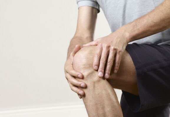
Pain syndrome in arthrosis
The pain is characteristic of arthrosis. They intensify with movement, physical activity, with changes in weather conditions, with changes in temperature, humidity level and atmospheric pressure. Pain can be triggered by any position of the body or sudden movements. Walking, running and prolonged vertical standing put some strain on the inflamed joints, after which a sharp or aching pain begins. In the first and second stages of the pathology, the pain syndrome disappears without a trace after a night's rest, but in an advanced stage, the pain is constant and does not disappear. The affected shock absorber layer, pinched nerves and blood vessels lead to the stagnation of the process with impaired trophism and accumulation of interstitial fluid. The swelling provokes a sharp throbbing pain.
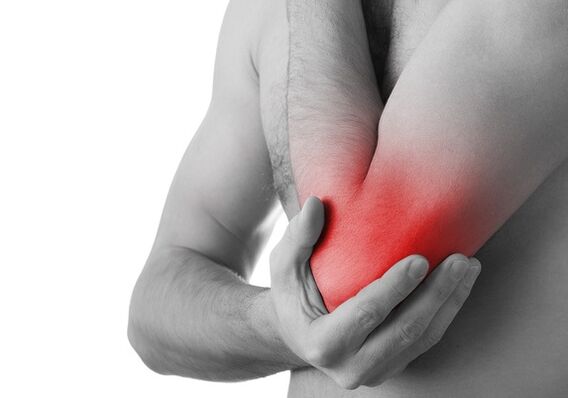
Specific for arthrosis is the pain after a long rest with a sharp motor impulse, this condition is called starting pain. The mechanism of development of these pains is osteophyte areas covered with destructive remnants of cartilage tissue, fibrin and viscous fluid. When the joints move, a film of these components or detritus covers the exposed areas, lubricating them and thus absorbing the pain. Blockade pain occurs after destruction products from the intra-articular space, i. e. bone debris or a large film of connective tissue, enter the muscles. There is another type of pain: constant, aching, popping + independent of movements, they are characteristic of reactive synovitis.
attention!The blocking type of pain is amenable only to surgical intervention, followed by restoration of the affected joint. Treatment with folk remedies is not recommended, this is fraught with the development of purulent arthrosis with the spread of infection in the body, and after sepsis there are obvious morphological changes in all organs and systems.
Symptoms of joint inflammation
Symptoms are divided depending on the degree of development of the pathology. Arthrosis is felt after 38-40 years, when the joint cushioning system begins to wear out and no renewed or young cartilage pads appear in its place. With a hormonal imbalance, "chaos" occurs in all vital systems, this also applies to the musculoskeletal system, so in the affected areas the tissues do not regenerate, but destruction + deformation occurs.
Symptoms of arthrosis:
| Degrees and periods of arthrosis | Description of symptoms |
|---|---|
| I degree |
|
| II degree |
|
| III degree |
|
| Periods of exacerbation and remission | In arthrosis, exacerbations alternate with remissions. The pathology is aggravated by physical activity. Exacerbations are caused by synovitis. The pain syndrome covers all affected areas, including the muscle corset. He reflexively spasms, forming painful contractures. Arthritis is characterized by muscle cramps. As the destruction increases, the pain syndrome becomes more pronounced. In reactive synovitis, the joint increases in size and acquires a spherical shape. A liquid appears in the joints, which creates a fluctuation effect when palpated. During a short remission, the pain subsides, but movement is difficult. |
Timely detection of the pathology with the help of diagnostic tests and consultation with the necessary specialists will help to pass the second and third stages, maintaining the functionality and health of all joint groups of the musculoskeletal system until old age.
Diagnostic measures
Clarification of the diagnosis is based on laboratory / instrumental studies. Each case is studied differently, that is, with an individual approach to each patient.
The research list consists of:
- General and biochemical blood tests.
- Blood test for rheumatoid agent.
- Analysis of urine and feces.
- X-ray examination: image in three positions.
- CT of the joint to clarify the bone structure.
- MRI of the joint: examination of ligaments and muscles.
- CT.
important!Patients with arthrosis need a consultation with an orthopedist, rheumatologist, endocrinologist, hematologist, oncologist, and women are recommended to consult a gynecologist.
Treatment regimen
Therapeutic tactics include a whole set of measures aimed at eliminating the main cause, adjusting the nutritional diet, restoring the lost function + a gentle lifestyle, that is, without special physical activity (prolonged walking, running, carrying heavy objects). The therapeutic treatment regimen consists of drug therapy, topical treatment, physical therapy procedures, and exercise therapy. In parallel with these methods, folk remedies are also used.

Drug therapy for arthrosis
Complex therapy consists of:
- Medicines from the group of NSAIDs;
- Painkillers (tablets + injections);
- Medicines that relieve muscle spasms (muscle relaxants);
- Cartilage tissue restorers (chondroprotectors);
- antibiotics;
- antihistamines;
- drugs that improve blood circulation;
- Vitamins: B2, B12, PP and A;
- Antioxidants: vitamin C;
- Medicines based on hormonal substances.
In the scheme of treatment of rheumatoid arthritis, it is recommended to include:
- Gold-based medicines;
- Immunosuppressants;
- Antimalarial drugs;
- Medicines that inhibit malignant cells.
attention!During remission of the pathology, non-steroidal anti-inflammatory drugs are not recommended, they affect the gastrointestinal tract, causing multiple ulcers, and also inhibit the process of nutrition of cartilage tissue.
Ointments for local application in arthrosis
Local treatment has a direct effect. Gels and ointments enter directly into the affected tissues, quickly reach the site, eliminating pain and inflammation. Preparations in the form of gels are widely used to restore the cartilage layer. For local application, warming + anti-inflammatory ointments are used.
Physiotherapy
Alleviation of spasmodic pain with reduction of inflammation + improvement of trophic and innervation is carried out with the help of physiotherapy. Exacerbation phases are eliminated or shortened by laser therapy, magnetic fields and ultraviolet irradiation. In the remission phase of arthrosis, i. e. during the quiescent phase, electrophoresis procedures with dimethylsulfoxide and anesthetics are useful. Destructive and inflammatory processes are affected by phonophoresis with glucocorticosteroids, inductothermy, heat applications of ozokerite or paraffin, as well as sulphide, radon and sea baths. The muscle corset is strengthened using electrical stimulation.
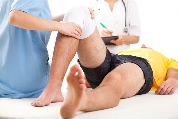
surgery
The problem of the deformed/ankylosed joint is finally solved by surgical operations such as endoprosthetics, as well as a palliative method of unloading the joint frame (coxarthrosis is eliminated by transtrochanteric osteotomy + femoral fascia fenestration; gonarthrosis is corrected by arthrotomy with cleaning of the intra-articular space from remnants ofdestruction plus artificial cartilage augmentation). If the bone is completely inoperable, it is replaced with an artificial graft and the axis of the tibia is corrected.
People's funds
Traditional medicine helps to get rid of pain and inflammation, temporarily removes pain and restores lost function. There are isolated cases of complete healing through traditional methods using the following tinctures, ointments and compresses:
- Tincture of garlic + onion and honey: 100 g of garlic mash + 100 g of chopped onion + 2 large spoons of honey + 200 ml of vodka. It is infused for 3-5 days. It is applied in the form of compresses and rubbing.
- Sabelnik in the form of tincture: 200 g of dry powder or fresh pulp + 200 ml of diluted medical alcohol, leave for 24 hours. Drink a spoonful before meals 3 times a day.
- Ointment based on badger fat and propolis: rub the joints, apply twice a day.
- Horseradish + honey meal: 100 g horseradish + 100 g honey + 100 ml vodka. Insist for 24 hours, drink 20 drops. With this tincture, you can smear the diseased joints 3-5 times a day.
- Hot pepper ointment + lard: 1 teaspoon powder + 200 g fat. It is infused for 2-3 days. It is used as a warming local medicine. Apply 1-2 per day.
- Compress: oak bark + spruce needles: 200 g oak bark + 200 g crushed spruce needles + 100 ml alcohol.
All listed recipes from folk healers are recommended to be used only after consulting a doctor. If the patient is allergic to certain drugs, their use is strictly prohibited, as they can lead to anaphylactic shock.
Features of prevention
Prevention is an effective tool to prevent joint diseases, destruction and deformation. For preventive purposes, you should do the following:
- Correct the menu, from which exclude fried, fatty, peppery, salty, alcohol + nicotine.
- Add jellies and jellies to your daily menu.
- Avoid tiring loads.
- Take precautions to avoid injury.
- Constantly perform a special set of exercises for the musculoskeletal system.
- Try to take vitamins B and C.
- For preventive purposes, take chondroprotectors, calcium, potassium and other mineral supplements once every six months.
- After spraining a joint or mechanical injury, get examined by a doctor.
The list is joined by performing constant physical exercises to improve blood supply, innervation and restoration of the cartilage layer of the joints. These exercises are prescribed by a doctor.
Summary
Destruction with deformation of the joints begins after 38-40 years, so it is not necessary to postpone the fight against this pathology. A neglected condition can lead to a wheelchair, and a timely response to the disease with effective treatment is a clear success towards recovery. It is impossible to treat arthrosis alone, this type of pathology refers to metabolic disorders directly related to changes in hormonal levels or chronic pathologies of other systems. At the first symptoms, contact a traumatologist or surgeon, do not delay, otherwise you will be treated only in a surgical department with long rehabilitation.